


Intervertebral disc herniation is more common in age-related osteochondrosis due to the dryness and fragility of the annulus fibrosus. But that is just one of the risk factors. Other include:
- High load in the lumbar region due to overweight.
- Weakness of the muscular system.
- Heredity.
- A sedentary lifestyle and, as a result, a constant compression of the vertebral structures.
- Smoking.
- Great physical activity.
According to medical statistics, this disease is often more common in men than women.
What are the most common causes of the disease:
- Injuries resulting from road accidents or falls.
- Lifting heavy objects with incorrect load distribution.
- Scoliosis or lordosis, which leads to increased stress in certain areas of the spine.
- Dysplasia of the hip joints.
- Chronic diseases including spinal tuberculosis, tumors, syphilis.
- Metabolic disorders (hereditary and acquired).
All of these factors cause wear and weakening of the cartilage and bones in the spine. And this is the main reason for the development of intervertebral hernia.
Stages of disease development
Without proper treatment, the disease progresses and the condition of the damaged spinal plates worsens. There are four stages in the development of the disease:
- Descent. The intervertebral disc was displaced quite a maximum of two millimeters. The nucleus pulposus does not extend beyond the vertebral body.
- Lumbar prominence. The edge of the disc extends beyond the vertebral body to 1. 5 mm, but no displacement of the nucleus is observed.
- Extrusion. The nucleus extends beyond the vertebral body.
- Sequestration. The nucleus virtually falls out and hangs in the form of a drop above its vertebrae. At this stage, there is a risk of cracking of the annulus fibrosus and leakage of fluid.
In the first stage of the disease, one is almost not worried about anything, sometimes there are back pains, but they go away quickly. With the development of the disease, the state of health also deteriorates, the symptoms become more painful and alarming. If the diagnosis is not made in time and treatment is not started, the consequences are possible: paralysis of the legs and severe disorders of the nervous system.
How does lumbosacral hernia manifest itself?
Intervertebral hernia can present with the following symptoms:
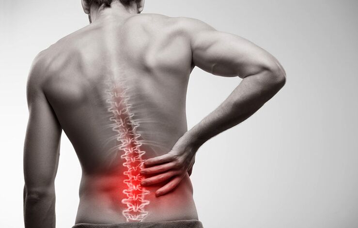
- Pain in the lumbar region.
- Pain while walking that radiates to the thigh region.
- Numbness of the surface of the feet, toes, legs and thighs.
- Feeling of difficulty in the legs.
- Stiffness of movement.
In order not to be late for medical help, it is worthwhile to analyze the symptoms of the disease more thoroughly. They can be divided into three groups.
Pain syndrome
Pain with lumbosacral spinal cord injury is a key symptom. Even in the first stage, there is pain in the area of the damaged disc, especially after injury. They may increase or decrease and then reappear. More often, the sacral region does not hurt either, but it hurts, especially during physical exertion or prolonged sedentary work. If a person lies on their healthy side and bends their legs, the pain will completely go away. This condition can take several months.
It is easy to get rid of the problem by treating medical help in a timely manner. It is enough to say goodbye to bad habits and perform the physiotherapy exercises recommended by your doctor.
Every day, the affected area grows and the condition of the disc tissue deteriorates. The transition to the second stage of the disease is indicated by increased pain. Today, it is not only felt in the sacral region, but covers the entire lower back, radiating to the neck region, individual spines, buttocks, thighs, legs, feet, and toes. The discomfort is manifested by physical activity, even insignificant - coughing or sneezing.
vertebral syndrome
The increased pain in the second stage is accompanied by constant cramps in the back muscles. This leads to even greater discomfort for the patient. He cannot move freely, straighten his back, stretch. The gait of such a person becomes precarious, he always leans to the opposite side of the patient, deteriorates.
The quality of human life is deteriorating due to impaired motor coordination. Unable to perform the tasks set at work well, active rest due to constant pain becomes unrealistic.
Radicular syndrome
If the hernia is left unattended by doctors, a progressive disease leads to compression of the roots of the spine, causing them to die and making it almost impossible for the tissues of the damaged disc to access the blood. Symptoms of severe stages of the disease appear:
- Weakening of the leg muscles. The patient cannot squat, stretch, jump. Even climbing stairs is hard for him.
- Numbness of the affected area and surrounding areas. The skin becomes numb and pale, with a goosebumps and tingling sensation. Patients complain of hyperhidrosis of the affected area and legs, or on the contrary, excessive dryness of the skin.
- Lumbago. The patient suffers from lumbago in the lumbar region, with acute, sharp pain that intensifies with each movement. If left untreated, it can lead to the destruction of the hip and knee joints.
- Noticeable thinning of the sore leg, leading to asymmetry of posture.
- Disruption of pelvic organs. Urological and gynecological diseases worsen, libido disappears, diarrhea, urinary incontinence are possible.
There is a risk of paralysis, disability and even death in the event of a severe spinal hernia.
Diagnosis of pathology
If a person has severe low back pain, they should make an appointment with a neurologist. You will be tested with medical tests:
- Identification of tendons reflexes in the lower extremities.
- Foot lift testing.
- Determination of heat or cold sensitivity, pain, and vibration over the entire surface of the legs, thighs, buttocks, abdomen, and back.
The doctor will then refer the patient for an MRI or CT scan of the lumbar spine. Tomographic techniques are used to obtain a three-dimensional image of the affected area. It can be used to determine the location and size of the hernia, the stage of the disease.
Electromyography, neurography, and contrast myelography are also prescribed if there is a risk of spinal cord injury. These tests help the doctor decide if urgent surgery is needed.
Treatment of disc herniation
Vertebral hernia is treated conservatively and surgically. The choice of technique depends on the stage of development of the disease, the presence of concomitant diseases and contraindications.
Conservative therapy
The therapeutic course is primarily aimed at relieving pain and relieving the patient’s condition.
What medicines can your doctor prescribe:
- Medicines to relieve pain and inflammation. In case of exacerbation - by injection. When the acute pain is relieved (usually three to four days is enough), oral medications with similar effects are prescribed.
- Novocaine blockade with the addition of corticosteroids. A similar method can stop the pain for two weeks at a time. Usually, the occlusion process is performed by injecting into different parts of the damaged plate.
- Centrally acting muscle relaxants. They reduce muscle activity by relieving pain cramps.
- Vitamin-mineral complexes with emphasis on Group B elements. They relax the muscles slightly, helping tissue to regenerate and conducting nerve impulses.
After pain syndrome is relieved, medication intake decreases. Treatment of the disease is the result of physiotherapy and physiotherapy.
Physiotherapy treatments are also selected depending on the condition of the patient. This could be:
- Treatment with heat or electric shock.
- Electrophoresis with anti-inflammatory drugs.
- Acupuncture and acupressure.
- Hirudotherapy.
- Hydromassage.
Normal massage is only allowed if there is no pain syndrome. More effective physiotherapy treatment is manual therapy with post-isometric relaxation.
Doctors strongly recommend that smokers quit smoking.
Dietary modification is also important, especially in overweight patients. Fatty, salty foods, sweets and alcohol should be excluded from the menu. A gentle diet with plenty of vegetables and fermented dairy products will help the body cope better with the treatment and get rid of the pounds that weigh on the back.
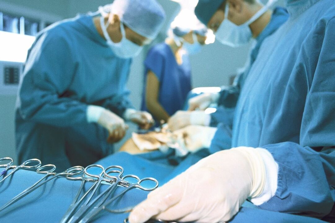
Surgical intervention
Conservative treatment usually lasts about two months. If you do not get the results you want, a decision is made to change your therapeutic tactics or perform the surgery. The latter is prescribed for severe pain, loss of sensitivity of the legs, dysfunction of the pelvic organs. Depending on the complexity of the situation, the operation is as follows:
- Endoscopic method. Three microsections are performed in the affected area. A camera is inserted into one to broadcast to the monitor. Through the other two, the protrusion of the hernia is removed with miniature instruments.
- By percutaneous discectomy. The damaged nucleus is removed by puncturing the disc and replaced with artificial material.
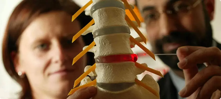
- With laser reconstruction. They are performed in the form of punctures, using a special needle, without separating the tissue. Laser radiation warms the disc structures and stimulates cell regeneration as well as relieves pain.
In severe cases, endoprosthesis of the vertebral plates is possible - replacement of the damaged organ with an implant.
Rehabilitation will be required after complex surgical procedures. The surgeon must wear a corset and will not be able to take a sitting position for about three months. The additional rehabilitation period includes the practice of therapeutic gymnastics and physiotherapy.
Preventive techniques
Like all other diseases, disc herniation is easier to prevent than to cure. What you need to do to keep your cartilage healthy:
- Calculate the exact loads if your work is related to them or if you are a professional athlete.
- Adequate body weight (index should not exceed 30).
- Choose a good mattress to sleep in the right position (preferably on your back).
- Participate in gentle physical education, swimming, fitness.
- Include exercises in the morning to strengthen the muscular corset of the spine.
- Quit the cigarette.
- Eat well.
If it becomes customary to follow these rules, there is a risk of spinal hernia only as a result of an accident.
Disc herniation has serious consequences and the treatment of advanced cases is very long. To avoid surgery and complications, you should see a neurologist if you have pain in your back.
Osteochondrosis
The term osteochondrosis itself comes from two words: osteo - bone and chondrue - cartilage. Simply put, it is the ossification of cartilage. Although this interpretation is fundamentally wrong. Some go even further in their delusions and are certain that osteochondrosis means the deposition of salts in the joints. Moreover, table salt is said to be consumed in large quantities.
Pathogenesis
In reality, everything happens a little differently. And harder. And table salt, if it plays any role in the development of osteochondrosis, is very indirect. Osteochondrosis is based on dystrophy and degeneration of articular cartilage. It is not a disease in itself, but a pathological process that can be observed almost everywhere where there is connective cartilage tissue.
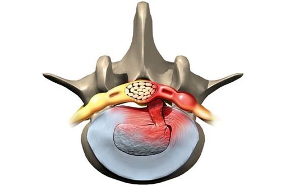
Nevertheless, osteochondrosis mainly affects the spine. Why is that? The situation is that there is a kind of spacer between the vertebrae - intervertebral (intervertebral) plates. The physiological role of these cartilage discs is to cushion and protect the vertebral bodies from premature wear due to mechanical stress. The disc consists of an inner liquid nucleus pulposus surrounded by an annulus fibrosus and an upper and lower endplate.
The plate is subjected to enormous mechanical stress, which leads to permanent damage to its cellular structures. In humans, these processes are too stressful - this is our pay for straight walking. In order for the disk not to be completely "erased", it must be continuously regenerated, i. e. restored. The balance of damage-regeneration processes determines the normal structure of the intervertebral disc. Another interesting detail is that the supply of blood and nutrients to the intervertebral discs is not through overgrown blood vessels in childhood, but in a diffuse way, from the bone tissue of the vertebral bodies. Again, the pay for the ability to walk on two legs is not four.
Because of this, intervertebral discs are easily damaged anatomically and physiologically. Any negative process in the body leads to imbalance of damage-regeneration, dystrophy and degeneration of the discs. A structurally defective disc can no longer withstand the proper mechanical stress. Excessive pressure on the lumbar vertebrae causes the cartilage discs to move in different directions, usually sideways and backward. This process is called disc herniation.
The bone tissue in the vertebrae that has lost its cartilage lining also goes through mechanical wear. Permanent trauma to the anterior edge of the vertebral bodies results in abnormal bone growth - osteophytes. Spondylosis develops. Due to the degeneration and displacement of the disc, the intervertebral spaces are reduced, the spinal canal is narrowed, and the roots of the spinal nerves are damaged. holes.
Cause
The causes or etiological factors of osteochondrosis are varied. Both can be local, e. g. caused by the pathology of the spine itself and general organizational abnormalities. Any pathology that leads to damage to the structure of the spine or metabolic disorders can be considered a cause of osteochondrosis. In this regard, they are:
- Changes in the configuration of the spine (scoliosis, pathological lordosis or kyphosis)
- Other defects of the musculoskeletal system - flat legs, narrow shoulder girdle, pelvic anomalies
- Spine injury
- Weak immunity
- Metabolic disorders - osteoporosis, obesity, diabetes mellitus, thyroid disease
- Cardiovascular diseases - atherosclerosis, high blood pressure
- Indigestion leading to insufficient absorption of nutrients from the gastrointestinal tract
- Heredity.
It should be noted that the above conditions do not necessarily lead to osteochondrosis. This requires constant exposure to certain predisposing factors - hypothermia, malnutrition, sedentary lifestyle or, on the contrary, excessive physical activity.
Symptoms
Osteochondrosis itself is an asymptomatic process. However, signs of intervertebral disc degeneration are varied. How come? The fact that the clinical manifestations of osteochondrosis are based on complications - disc herniation, spondylosis, sciatica, spinal stenosis.
In addition, the clinic varies greatly depending on whether there is a dominant localization of the process in the cervical, thoracic, or lumbosacral spine. The last stage is most often affected, as the lower back absorbs maximum physical activity. Signs of osteochondrosis of the lumbosacral region:
- Pain (lumbodynia, lumbago, sciatica)
- Restriction of movement in the lower back and lower extremities (intermittent hiccups)
- Sensitivity disorders of the type of paresthesia here - numbness, burning, crawling
- Abnormal tension in the lumbar muscles
- Disorders of the pelvic organs in the absence of treatment.
Cervical osteochondrosis is somewhat less common than lumbosacral. However, this pathology is quite common. In addition to the characteristic symptoms of pain (cervicalgia), decreased sensitivity, and upper limb movements, cervical osteochondrosis due to impaired blood supply to the brain also has its own characteristics. These characteristics are manifested:
- Insomnia
- Headache, dizziness
- Intermittent nausea
- General weakness, rapid fatigue
- Fluctuations in blood pressure
- Occasionally a toothache
- Behavioral reactions in the form of tearing and irritability.
The chest region of osteochondrosis is relatively rarely affected. Patients in this case are people who sit in a fixed, uncomfortable position because of their occupation - students, schoolchildren, programmers, office workers. Symptoms of osteochondrosis in this case include:
- Chest pain and paraesthesia
- Shortness of breath
- Feeling of a heartbeat
- Restriction of movement in the thoracic spine.
Diagnostics
From all this, it is clear that osteochondrosis is a chameleon disease. Due to the similarity of symptoms, it can be easily confused with cerebrovascular accident, hypertension, myocardial infarction, angina pectoris, neurotic disorders. Therefore, a comprehensive complex diagnosis is needed to establish the correct diagnosis in order to correctly define the symptoms and treatment of osteochondrosis.
In addition to traditional questioning and clarification of patient complaints, this diagnosis should include medical examination and special research methods. These methods include x-rays of the spine, ultrasound of the internal organs. Recently, computer and magnetic resonance imaging have been successfully used to diagnose osteochondrosis.
Treatment
Therapeutic tactics for osteochondrosis include:
- Medicines
- Massage
- Physiotherapy procedures
- Physiotherapy
- Manual therapy
- Acupuncture.
Medication for osteochondrosis is primarily aimed at relieving pain and eliminating inflammatory processes in the nerve roots. NSAIDs are used for this purpose. In various combinations, these drugs are widely used in the form of ointments, injections, tablets for the treatment of osteochondrosis. It should be borne in mind that these drugs have a negative effect on the liver, stomach and intestines. This can exacerbate metabolic disorders in osteochondrosis. Local anesthetics are good for relieving the pain of blockade. True, the effects of these funds are short-lived and do not affect osteochondrosis as a whole in any way.
Metabolic processes can be improved locally and at the body level with the help of drugs such as chondroprotectors, immunostimulants, vitamins with minerals. Chondroprotectors are used in tablets, ointments and ampoules. Strengtheners include vitamin C, group B, in combination with minerals. In this regard, calcium formulations are most preferred. In fact, contrary to some misconceptions, the basis of osteochondrosis is not an excess of calcium but merely a lack of it.
After successful relief of exacerbation, physiotherapy, massage and gym therapy will be introduced. Physical methods include calcium electrophoresis, hydrocortisone phonophoresis, amplification, and paraffin therapy. All of these measures are aimed at eliminating pain and inflammation in the nerve roots, ligaments, and muscles. Massage for osteochondrosis is performed according to a generally accepted method. The massage zone is selected depending on the localization of the osteochondrosis. The range of motion is expanded with the help of movement therapy. Initially, there is virtually no dynamic load in the exacerbation phase. The patient is in an optimal posture at all times. At this time it is desirable to wear fastening devices - lumbar corset, Shants collar. As exacerbation ceases, the amount and duration of movement during training therapy increases.
Recently, non-traditional methods of treatment have been used to treat osteochondrosis - acupuncture, manual therapy, osteopathy. Acupuncture affects specific biologically active points located along the spine, earlobes, hands, and feet. With manual therapy, the normal position of the vertebrae and intervertebral discs is restored by manual intervention by the hands of a specialist. In osteopathy, the structural integrity of the musculoskeletal system is ensured by specific techniques. In the absence of the effect of conservative measures for the treatment of osteochondrosis, lasting pain, complications and surgery are required. The abnormally displaced plate is removed. A microdiscectomy is currently being performed for this purpose - endoscopic removal of the displaced plate.